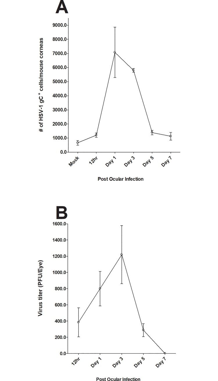
Fig 2. Monitoring the presence of virus in cornea of infected mice.
Mice were ocularly infected with HSV-1 as described above and viral titers were measured in the eye of infected mice by plaque assay 12h, 1, 3, 5 and 7 days PI using eye swabs from 20 eyes. At the indicated time points, corneas of infected mice were isolated and single cell suspensions were prepared as described in Fig 1 above. Single cell suspension was stained with FITC anti-gC antibody. Each point represents mean cell number ± SEM per 3 mice corneas. Experiments were repeated three times. Panels: A) gC expression in cornea of infected mice: and B) Virus titer in the eye of infected mice.

