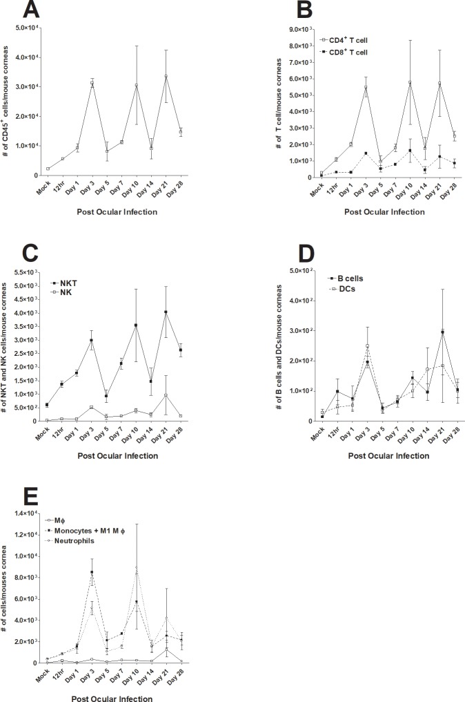
Fig 3. Effect of ocular HSV-1 infection on alteration of various immune infiltrates in cornea of infected mice at different times PI.
Mice were ocularly infected with 2 X 10⁵ PFU/eye of HSV-1 strain McKrae. Corneas of infected mice were harvested at days 12 h, 1, 3, 5, 7, 10, 14, 21 and 28 days PI. Single cell suspension of corneal cells was prepared and subjected to flow cytometry as described in Materials and Methods. Panels show the changes in the number of different macrophage subtypes at different times PI. Uninfected mice were used as mock control. For each time point we used a separate mock correspondent to the time that we performed the FACS analyses and due to their similarity in all tested time points, we only showed one mock in each figure. Each point represents mean cell number ± SEM per 3 mice corneas. Experiments were repeated three times. Panels: A) CD45+ immune cells; B) CD4+ and CD8+ T cells; C) CD4+ to CD8+ T cell ratio; D) NK and NKT cells; E) B cells and DCs; and F) F4/80+Ly6C- macrophages, F4/80+Ly6C+ monocytes and M1 macrophages, and F4/80-Ly6C+ neutrophils.

