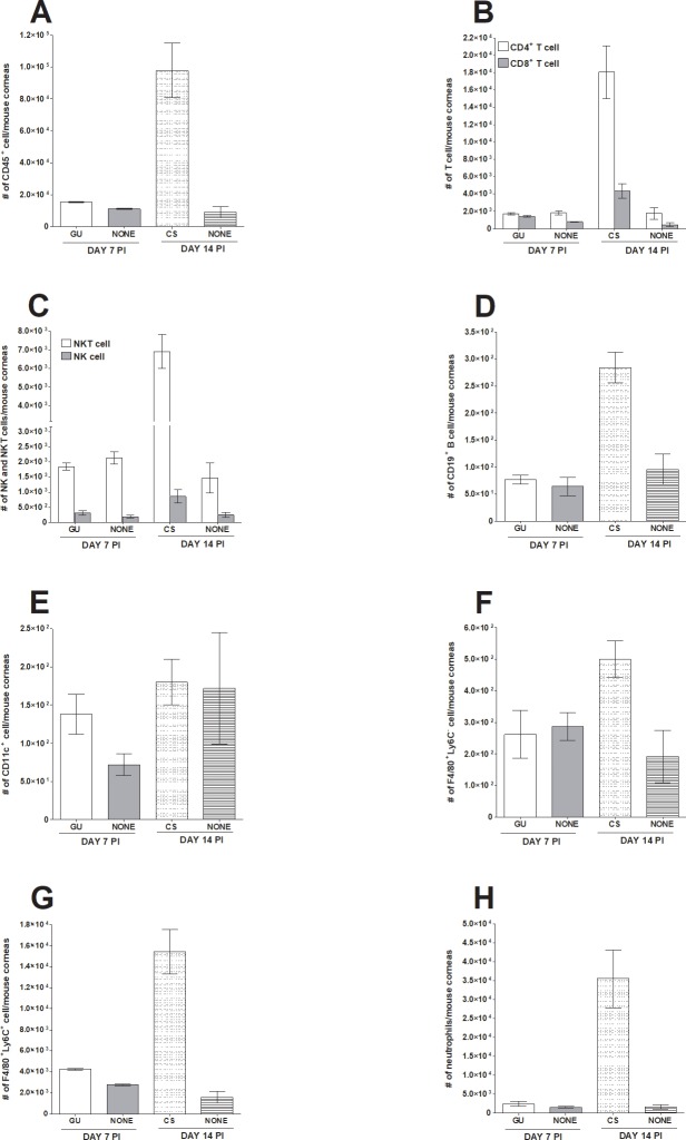
Fig 5. Comparison of infiltrating immune cells in the eye of infected mice with and without eye disease.
Mice were ocularly infected as described in Fig 1 above. On day 7 and 14 PI, mice showing geographical ulcer (GU) or corneal scarring (CS), respectively were euthanized, corneas were collected, and subjected to flow cytometry and compared with mice that were similarly infected but did not show any eye disease (none). Each bar represents mean cell number ± SEM from 3 mice corneas. Experiments were repeated three times. Panels: A) CD45+ immune cells; B) CD4+ and CD8+ T cells; C) NK and NKT cells; D) CD19+ B cells; E) CD11c+ DCs; F) F4/80+Ly6C- macrophages; G) F4/80+Ly6C+ monocytes and M1 macrophages; and H) Neutrophils.

