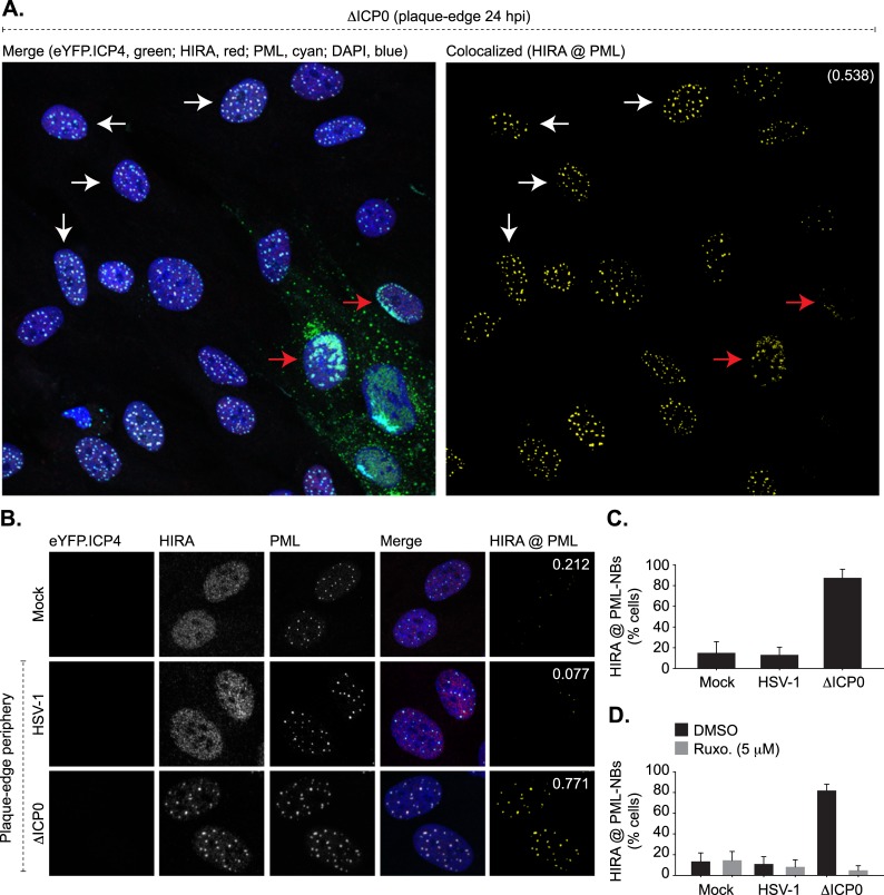
Fig 2. HSV-1 induced activation of innate immune signalling promotes HIRA localization at PML-NBs in a JAK-dependent manner.
HFt cells were mock infected or infected with 0.001 or 2 PFU/cell of WT or ΔICP0 HSV-1 recombinant viruses expressing eYFP.ICP4, respectively, to enable plaque-formation to occur. Infected cell monolayers were fixed and permeabilized at 24 hpi, and the nuclear localization of HIRA (red) and PML (cyan) were evaluated by indirect immunofluorescence. Nuclei were stained with DAPI (blue). (A) Wide-field plaque-edge image showing the nuclear localization of HIRA and PML in cells in close proximity to a developing ΔICP0 plaque-edge (eYFP.ICP4, green). Corresponding wide-field images for mock and HSV-1 infected cells are shown in S2 Fig. Cut mask (yellow) highlights regions of colocalization between HIRA and PML. Weighted colocalization coefficient is shown. Arrows highlight examples of cells that are either positive (red) or negative (white) for eYFP.ICP4 expression. (B) Nuclear localization of HIRA and PML in mock or eYFP.ICP4 negative cells at the periphery of developing WT or ΔICP0 plaque-edges (equivalent to white arrows in A). (C) Quantitation of the population of eYFP.ICP4 negative cells showing HIRA colocalization at PML-NBs (as shown in A, B, and S2 Fig). n ≥ 500 cells per sample population derived from four independent infections. Means and standard deviations (SD) shown. (D) Quantitation of the population of eYFP.ICP4 negative cells positive for HIRA colocalization at PML-NBs in mock, WT, or ΔICP0 HSV-1 infected cells treated with DMSO or the JAK inhibitor Ruxolitinib (Ruxo; 5 μM). n ≥ 500 cells per sample population derived from four independent infections. Means and SD shown. Wide-field images for ΔICP0 HSV-1 infected and DMSO or Ruxo treated cell monolayers are shown in S4 Fig.

