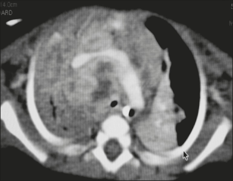
Figure 5.
Pulmonary tuberculosis presenting as a pseudotumor in a threemonth-old infant. Contrast-enhanced CT, with soft-tissue window settings, showing enlarged hypodense lymph nodes-pretracheal, right paratracheal, and caval-aortic-with expansile features, forming a mass that partially compressed the right primary bronchus. Note the consolidations in the upper lobes and in the apical segment of the left lower lobe.

