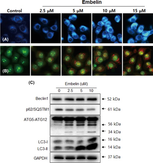
Figure 4.

Embelin induced autophagy in Ca9–22 cells. Cells were stained with MDC (A) and acridine orange (B) as described in Materials and Methods. Ca9–22 cells were grown on coverslips and treated with Embelin (2.5–15 μM). Autophagic vacuoles were observed and imaged on a fluorescence microscope. C, Cells were treated with various concentrations of Embelin for 24 h and the expression levels of autophagy‐related proteins, such as Beclin‐1, p62/SQSTM1, ATG5‐ATG12, and LC3, were analyzed by Western blotting. [Color figure can be viewed at http://wileyonlinelibrary.com]
