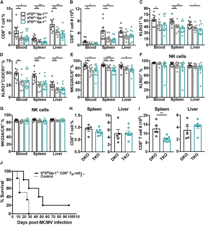
Figure 7. Loss of Qa-1 Negatively Impacts the Non-classical CD8+ T Cell Response and Their Protective Capabilities.
(A and B) Frequency (A) and absolute number (B) of non-classical CD8+ T cells in the spleen, liver, and blood of KbDb−/−Qa-1+/+, KbDb−/−Qa-1+/−, and KbDb−/−Qa-1−/− mice on day 7 post-MCMV infection (n = 9–11).
(C–G) KLRG1+CD127− (C), KLRG1+CX3CR1+ (D), and NKG2A/C/E+ (E) non-classical CD8+ T cells and KLRG1+ (F) and NKG2A/C/E+ NK (G) cells on day 7 post-MCMV infection from indicated organs of KbDb−/−Qa1+/+, KbDb−/−Qa1+/−, and KbDb−/−Qa1−/− mice (n = 9–11).
(H and I) Frequency (H) and absolute number (I) of CD8+ T cells from the spleen and liver of long-term infected KbDb−/−Qa1+/+ (DKO) and KbDb−/−Qa1−/− (TKO) mice (n = 5–7).
(J) Survival of RAG1−/−KbDb−/− mice with (—, n = 5) or without (- -, n = 6) adoptive transfer of 5 × 10⁴ sorted CD8+ TM cells (KLRG1CD27+) from long-term infected KbDb−/−Qa-1−/− mice. Statistical analysis determined by Mantel-Cox test.
Data are pooled from two (H and I) or three independent experiments (A–G) or representative of three independent experiments (J). Data represent mean ± SEM. *p < 0.05, **p < 0.01, ***p < 0.001, and ****p < 0.0001.
See also Figure S4.

