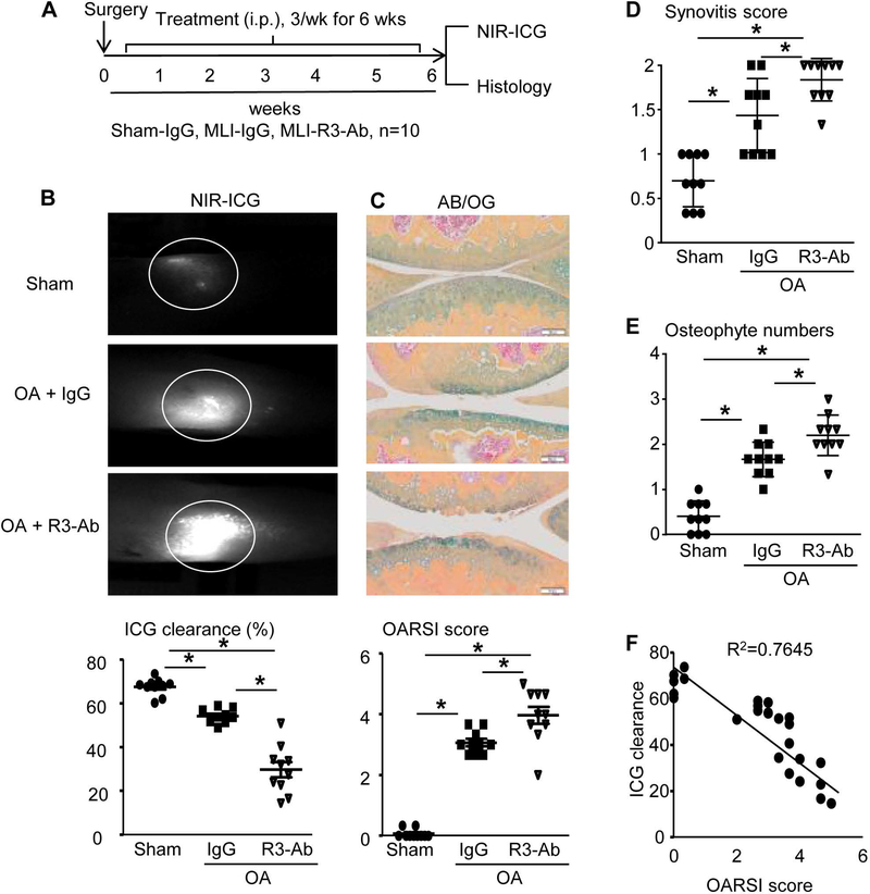
Figure 1. Inhibition of lymphangiogenesis via VEGFR3 blockade exacerbates cartilage destruction in PTOA joints.
(A) Schematic illustration of the experimental design in which WT C57BL/6J mice received MLI or sham surgery, and on day 3 post-surgery were randomized to treatment with anti-VEGFR3 neutralizing antibody (R3-Ab, 0.8 mg/kg, i.p. 3 times/week) or IgG (placebo) for 6 weeks. (B) Mice were subjected to NIR-ICG imaging to quantify synovial lymphatic drainage, and representative images of the ICG remaining in the knee 24hr post-injection (circled region) are shown to illustrate the lack of lymphatic clearance in OA + R3-Ab treated mice. (C) Paraffin sections of knee joints were stained with AB/OG for OARSI scores, and representative micrographs are shown at 10× (bar = 0.1mm). The synovitis score (D) and osteophyte numbers (E) are presented as the mean +/− SD, n=10 mice per group (*p<0.05 via One-way ANOVA followed by Tukey). (F) The relationship between ICG clearance and OARSI score was determined by linear regression analysis (p<0.05 via Pearson’s correlation coefficient; n=30).

