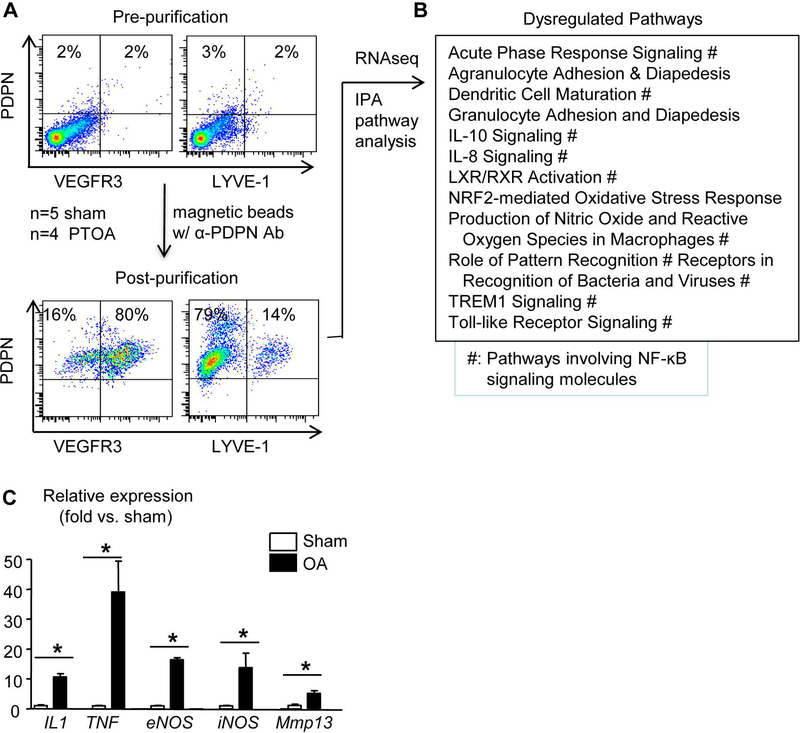
Figure 2. Lymphatic endothelial cells in PTOA synovium have an inflammatory phenotype.
Mice received MLI or sham surgery as in Fig.1. (A) LECs were isolated from synovium at 6 weeks post-surgery with anti-PDPN antibody, and the enrichment of this PDPN+ population was confirmed by flow cytometry (n=6 mice). (B) RNAseq was performed on the PDPN+ cells, which revealed about 1,000 differentially expressed genes in PTOA vs. sham synovial LECs. Pathway analysis indicated 12 dysregulated pathways (# indicates pathways involving NF-κB signaling; n=4–5 mice/group). (C) Synovial LECs from a different cohort of PTOA mice at 6 weeks post-surgery were subjected to qPCR. Data are mean + SD in which the Sham expression level = 1 (n=5 mice/group; *p<0.05 via unpaired t test).

