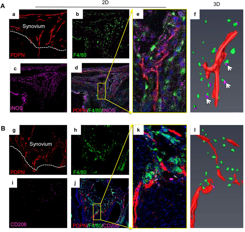
Figure 4. M1 macrophage accumulation adjacent to lymphatic vessels in PTOA synovium.
Frozen sections (30 μm thick) of knees at 5 weeks post-MLI were immuno-stained with antibodies against PDPN (red) for lymphatic vessels (a), F4/80 (green) for pan macrophages (b), iNOS or CD206 (purple) for macrophage subsets (c). M1s (A) were defined as F4/80/iNOS+ (a-d), and M2s (B) were defined as F4/80/CD206+ cells (g-l). Confocal microscopy was used for z-section imaging to obtain 20–25 consecutive images (e and k) with a step-width of 1μm. PDPN+ lymphatic vessels (red) and M1s or M2s (purple) were detected by Amira to generate 3D images (f and l) in a SurfaceGen module. Arrows in f indicate M1 cells near lymphatic vessels in a 3D image (n=4 mice).

