
Figure 3.
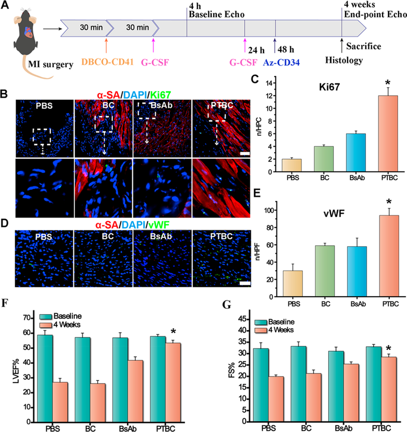
PTBC treatment promotes angiomyogenesis and boosts cardiac function in mice with MI. (A) Schematic showing animal study design. (B) Images showing Ki67-positive cardiomyocyte nuclei in control PBS-, DBCO-PEG-IgG followed with Az-PEG-CD34 (BC), BsAb or DBCO-PEG-IgG followed with Az-PEG-CD34 (PTBC)-treated hearts. (C) Quantitative analysis of Ki67-positive nuclei. (D) Representative micrographs showing vWF-tagged vessels (green) treated by control PBS-, DBCO-PEG-IgG followed with Az-PEG-CD34 (BC), BsAb or DBCO-PEG-IgG followed with Az-PEG-CD34 (PTBC)-treated hearts at 4 weeks. (F, G) LVEFs and LVFSs measured by echo at baseline (4 h post-MI) and 4 weeks later (n = 4 animals per group). All data are mean ± SD. Scale bar, 40 μm. PTBC group vs the other three groups; * indicates p < 0.05.