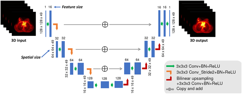
Fig. 1:
The schematic diagram of the neural network architecture. The feature size after the first convolution is 16. Once the image is downsampled through the convolution operation with stride 2, the feature size will be multiplied by 2. The spatial size is based on the XCAT and lung patient studies. For brain patient study, the third dimension of the spatial size is 91. 3D convolution is used and the total number of trainable parameters is around 1.4 million.

