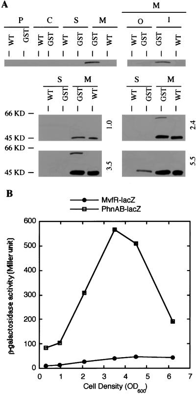
Figure 4.
(A) Cellular localization of the MvfR protein at different growth phases. Plasmid containing the MvfR-GST translational fusion was introduced into PA14 strain. Protein extracts from cell fractionations of both PA14 and transformant strains grown to cell density as indicated were prepared, separated on a 10% polyacrylamide gel, and blotted onto Immobilon-P [poly(vinylidene difluoride) (PVDF)] membranes. A mAb against GST was used to detect the MvfR-GST fusion. The numbers to the right indicate the cell density (OD600). WT, wild-type PA14 strain; GST, PA14 strain containing MvfR-GST translational fusion; P, periplasmic; C, cytoplasmic; S, secreted; M, membrane; O, outer; I, inner. (B) The expression of mvfR and phnAB at different growth phases. β-galactosidase activities in PA14 strain containing either mvfR-lacZ or phnAB-lacZ transcriptional fusion were measured at the growth phases as indicated on the graph.

