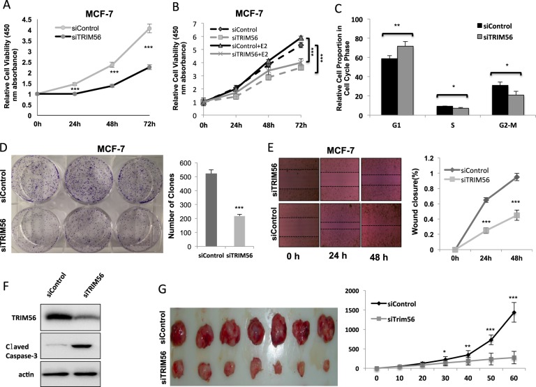
Fig. 2. TRIM56 depletion inhibits ER-alpha-positive breast cancer cell proliferation in vitro and in vivo.
A TRIM56 depletion inhibits the cell proliferation in breast cancer cells. MCF-7 cells were transfected with 50 nM TRIM56 siRNA (mix of #1 and #2) or 50 nM control siRNA. After 24 h, the WST assay was used to determine the cellular metabolic activity at indicated time points after transfection. Experiments were done in triplicates. *P < 0.05; **P < 0.01; ***P < 0.001 for cell growth comparison. B TRIM56 depletion inhibits estrogen-driven cell proliferation in breast cancer cells. MCF-7 cells were transfected with 50 nM TRIM56 siRNA (mix of #1 and #2) or 50 nM control siRNA. After 24 h, cells were treated with vehicle or 10 nM estradiol. The WST assay was used to determine the cellular metabolic activity at indicated time points after transfection. Experiments were done in triplicates. *P < 0.05; **P < 0.01; ***P < 0.001 for cell growth comparison. C TRIM56 depletion significantly induces G1 cell cycle arrest in breast cancer cells. MCF-7 cells were transfected with 50 nM TRIM56 siRNA (mix of #1 and #2) or 50 nM control siRNA. After 48 h, cells were harvested and fixed by 70% ethanol. The cell cycle phase was anaylzed by PI staining. Experiments were done in triplicates. *P < 0.05; **P < 0.01; ***P < 0.001 for cell growth comparison. D Clone formation assay of MCF-7 cells transfected with indicated 50 nM TRIM56 siRNA (mix of #1 and #2) or 50 nM control siRNA. Quantification of clone formation is shown at the indicated time points. Data are presented as ±SD. **P < 0.01, ***P < 0.001 (Student’s t test). E Wound-healing assay of MCF-7 were transfected with indicated 50 nM TRIM56 siRNA (mix of #1 and #2) or 50 nM control siRNA. Quantification of wound closure at the indicated time points. Data are presented as ±SD. **P < 0.01, ***P < 0.001 (Student’s t test). F TRIM56 depletion promotes apoptotic signaling in breast cancer cells. T47D cells were transfected with indicated 50 nM TRIM56 siRNA (mix of #1 and #2) or 50 nM control siRNA. After 24 h, cells were harvested for western blot analysis. The cleaved caspase-3 was detected to indicate apoptosis signaling activity. G TRIM56 depletion inhibits the cell proliferation in breast cancer cells in vivo. MCF-7 cells were stably transfected with lentivirus carrying a scrambled shRNA or TRIM56 shRNA. Female NOD scid gamma (NSG) mice were estrogen-supplemented by implantation of slow-release 17β-estradiol pellets (0.72 mg/90-d release; Innovative Research of America) 1 day before MCF-7 tumor cell injection into the mammary fat pad (2 × 10⁶ MCF-7 cells suspended in 100 μl Matrigel solution). MCF-7 tumor xenografts were measured every 10 days and the tumor volume was calculated by length × width2 /2. The mice were killed 2 months after transplant. The tumor growth curve and a photograph were shown

