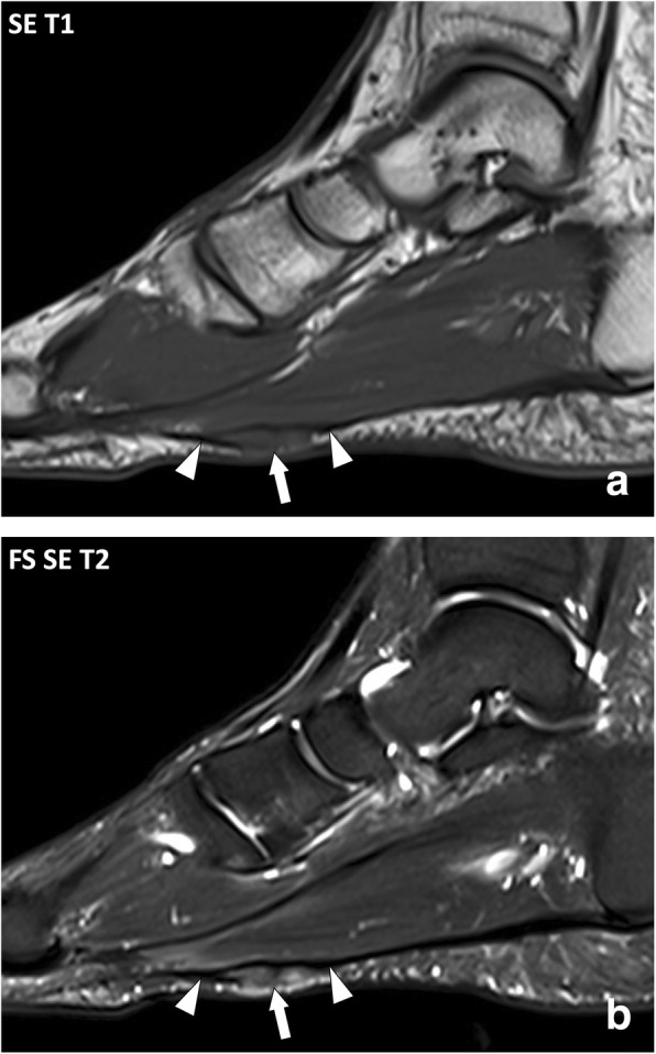
Fig. 13.

Sagittal (a) SE T1w and (b) fat-suppressed SE T2w images of the right foot of a 47-year-old male with plantar fibromatosis. MRI demonstrates fusiform thickening of the plantar fibromatosis with a nodule of low signal intensity on the T1w image and heterogeneous high signal intensity on the T2w image (arrow) in continuity with the normal aponeurosis (arrowheads)
