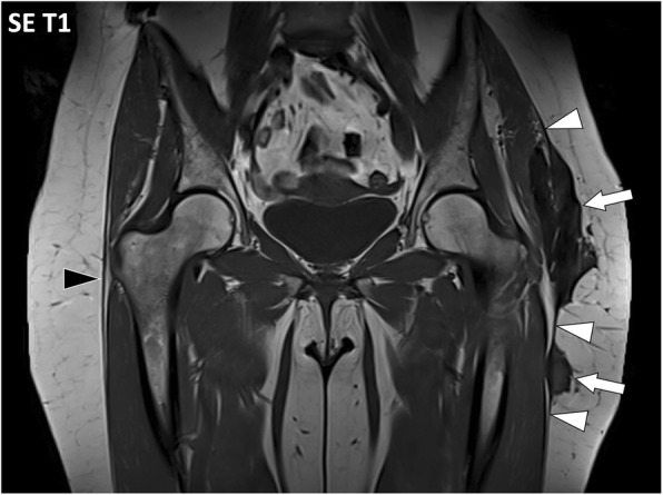
Fig. 14.

Coronal SE T1w image of the pelvis of a 52-year-old female with a desmoid tumor in the left hip area. MRI demonstrates low signal intensity masses (arrows) in continuity with the iliotibial tract and the deep peripheral fascia with typical aspect described as “fascia tail sign” (white arrowheads). The right iliotibial tract and deep peripheral fasciae are normal (black arrowhead)
