
Fig. 16.
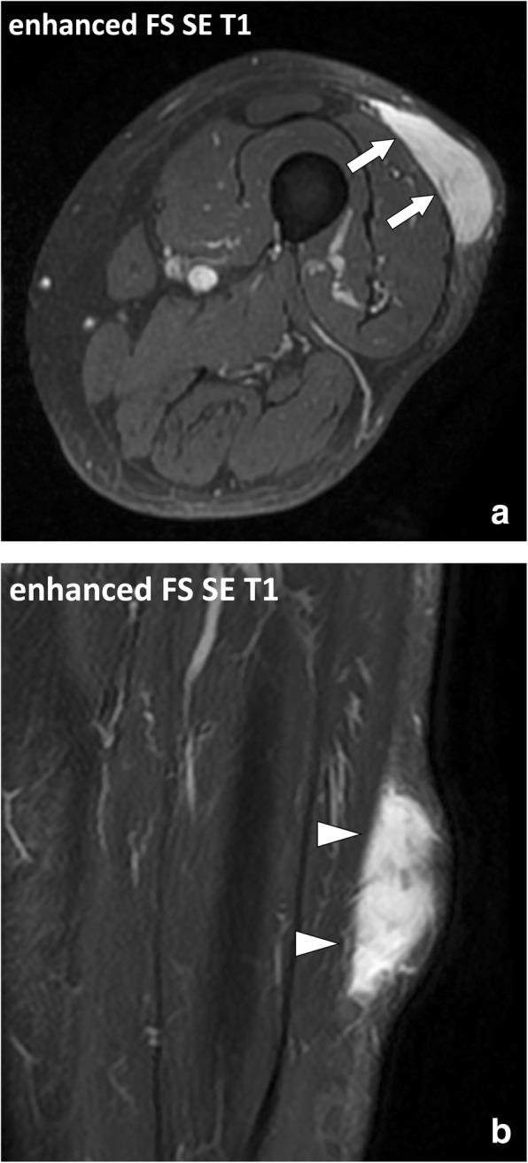
Focused (a) axial, i.e., short axis, and (b) coronal, i.e., long axis, SE T1w contrast-enhanced fat-suppressed images of the left thigh of a 75-year-old male with high-grade myxofibrosarcoma. Axial images clearly demonstrate the localization of the lesion in the hypodermis and its anatomical relationship with the underlying deep peripheral fascia (arrows). The deep fascia is less conspicuous on the longitudinal images due to partial volumes (arrowheads)