
Fig. 2.
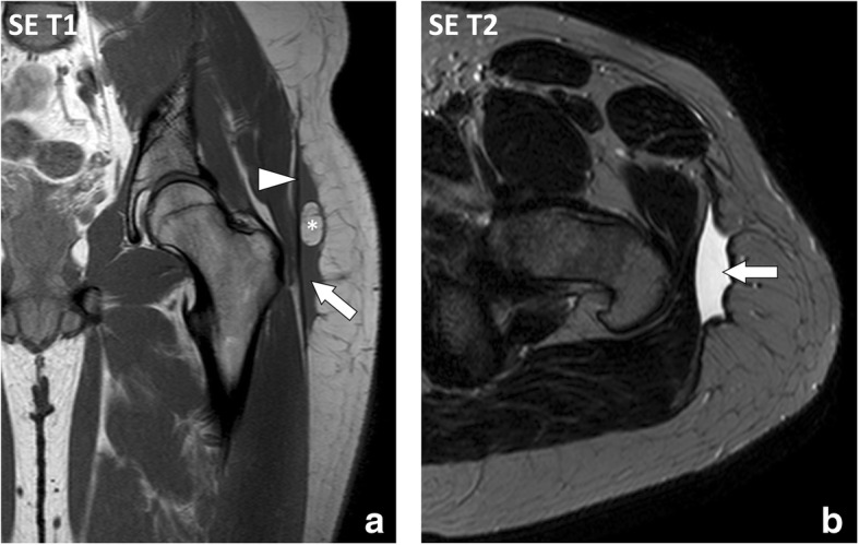
a Coronal SE T1-weighted (T1w) and (b) axial SE T2-weighted (T2w) images of the left hip and proximal part of the left thigh of a 22-year-old male with Morel-Lavallée lesion after a street fight. MRI demonstrates an extensive lenticular fluid collection (arrows) deep to the hypodermis and superficial to the fascia lata (arrowhead). Note the large fat lobule bulging into the collection (asterisk)