
Fig. 3.
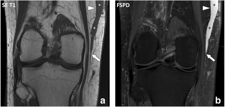
Coronal (a) SE T1w and (b) fat-suppressed proton density-weighted (FSPD) images of the right knee of a 23-year-old male with Morel-Lavallée lesion after a motorcycle accident. MRI demonstrates a fluid collection (asterisks) extending from the interface between the hypodermis and deep peripheral fascia (arrow) along the fascia superficialis (arrowhead)