Fig. 9.

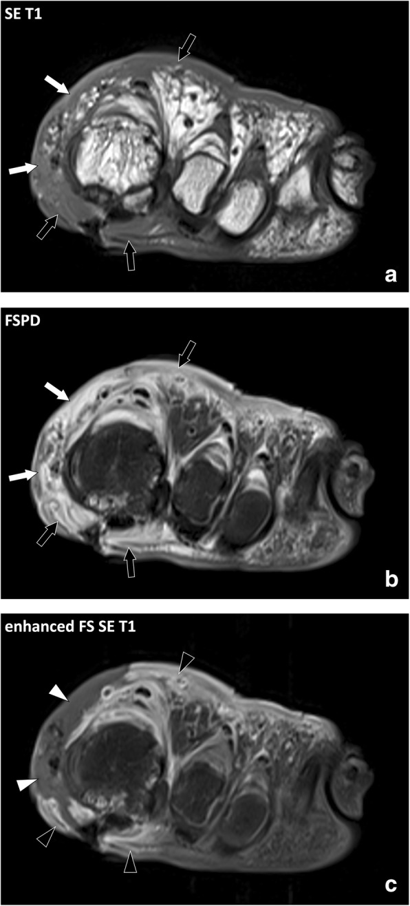
Coronal (a) SE T1w, (b) FSPD, and (c) contrast-enhanced fat-suppressed T1w images of the left forefoot of an 81-year-old diabetic male with foot ulcer and necrotizing cellulitis. SE T1w and fluid-sensitive images demonstrate infiltrated hypodermic fat on the medial (white arrows) and to a lesser account dorsal aspect (black arrows) of the foot while the hypodermic fat on the plantar and lateral aspect is normal. After contrast material injection, necrotized skin and fat do not enhance (white arrowheads) and are surrounded by fat with enhanced inflammatory infiltration (black arrowheads)
