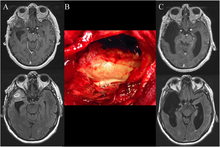
Figure 1.
(A) Preoperative axial enhanced T1-weighted MRI (upper) and FLAIR-weighted MRI (lower) achieved in a 50-year-old right-handed man who experienced seizures that allowed the discovery of a right anterior temporal high-grade glioma. The patient underwent a previous “minimal-invasive image-guided surgery” performed under general anesthesia in another hospital with a partial resection of the enhancement and the FLAIR hypersignal. An anaplastic astrocytoma was diagnosed. At that time, the patient was referred to our department and a reoperation was proposed with awake mapping in order to achieve a supratotal resection according to functional boundaries. The neurological examination was normal. Nonetheless, the preoperative neuropsychological evaluation revealed a slight deficit of higher-cognitive functions, that is, theory of mind and semantic processing. (B) Intraoperative view after resection, achieved up to eloquent structures, especially at the subcortical level. Indeed, direct electrostimulation of white matter tracts enabled the identification of the subcortical neural networks involved in theory of mind (mentalizing) (tag 47) and non-verbal semantics (tag 49) - which have been mapped according to the results of the presurgical neurocognitive assessment. (C) Postoperative axial enhanced T1-weighted MRI (upper) and FLAIR-weighted MRI (lower) (performed 3 months after resection) demonstrating a supracomplete resection of both the enhancement and the FLAIR hypersignal. The patient recovered, with an improvement of the neuropsychological examination thanks to a post-surgical cognitive rehabilitation. A diffuse WHO grade III astrocytoma (IDH1 mutated, non-codeleted) was diagnosed, and postoperative chemotherapy was administrated, with no radiotherapy. The imaging is stable with 4 years of follow-up, and the patient continues to enjoy a normal life, with no symptoms.

