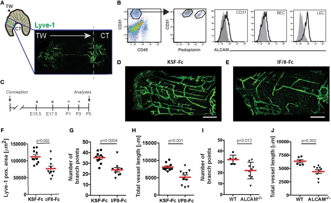
Figure 3.
I/F8-Fc treatment reduces developmental lymphangiogenesis in the diaphragm to a similar degree as ALCAM deficiency. (A) Schematic representation of the diaphragm and of the area analyzed by LYVE-1 immunofluorescence in D-J. TW: thoracic wall; CT: central tendon. (B) ALCAM expression on diaphragmatic P5 BECs (CD45−CD31+podoplanin−) and LECs (CD45−CD31+podoplanin+) was confirmed by FACS. Data from 1 out of 2 similar experiments are shown. Specific stainings are shown as black lined and isotype control stainings as gray tinted histograms. (C) Schematic presentation of the antibody treatment protocol. Pregnant mice received I/F8-Fc or KSF-Fc antibody i.p. (300 μg) on day E15.5 and E17.5 (indicated by an asterisk). In addition, newborn mice received antibody i.p. (30 μg) on day P1 and P3 (indicated by a circle). (D–H) On P5 the diaphragm was collected and lymphatic vessels were visualized by LYVE-1 staining. (D,E) Representative stainings from diaphragms of KSF-Fc- or I/F8-Fc-treated animals. (F–H) Image-based morphometric analysis of (F) the LYVE-1+ area, (G) the number of branch points and (H) the total vessel length in the diaphragm of I/F8-Fc or KSF-Fc control-treated pups. Each dot represents the measurement made in one animal (n = 10). Data from 1 out of 3 similar experiments are shown in F-H. (I,J) Image-based morphometric analysis of (I) the number of branch points and (J) the total vessel length in the P5 diaphragm of WT and ALCAM−/− mice. Data from 1 experiment (n = 6 or 9 mice) are shown in G-H. KSF-Fc: control antibody. I/F8-Fc: anti-ALCAM.

