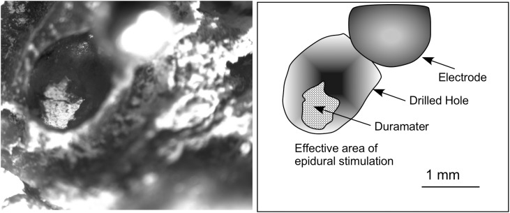
FIGURE 2.
Superficial view of the skull after post-mortem electrode extraction (the electrode was twisted and is shown in the figure close to the drill bit on the right-hand corner, out of focus). Glued to a coverglass, the bone was placed in the microscope stage and transilluminated to take the picture. The contour of the surface of dura mater exposed to the electrode is clearly shown using this approach. Measures of the surface were used to correlate with EES effects on the brain surface and to estimate the amount of electric current injection.

