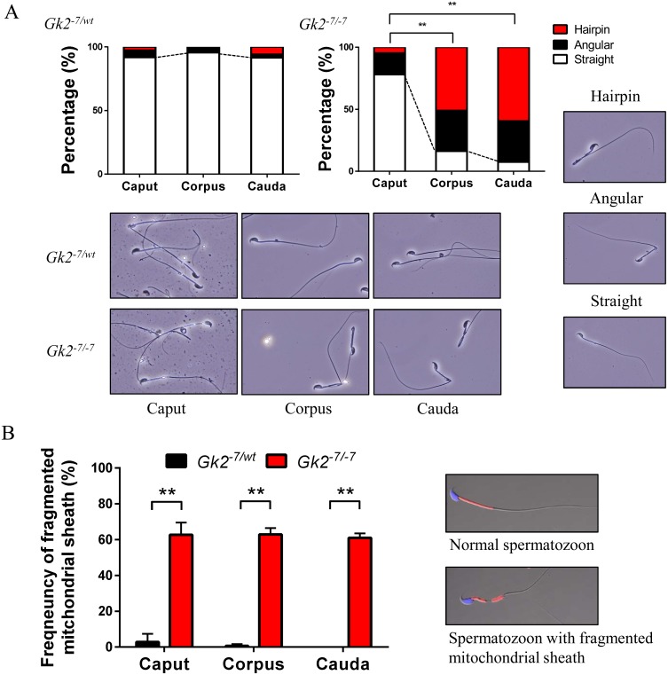
Fig. 2.
Mitochondrial disorganization of Gk2 disrupted spermatozoa appears before sperm bending. (A) Graphs indicate frequencies of bending spermatozoa collected from the three sections of epididymis. Lower panels show representative images of spermatozoa collected from the three sections of epididymis. Spermatozoon shape is categorized as Hairpin, Angular, or Straight (displayed in right panels). ** P < 0.01, Student’s t test. (B) Graph indicates the frequencies of fragmented mitochondrial sheath collected from the three sections of epididymis. Right panels are representative images of spermatozoon with or without a fragmented mitochondrial sheath. ** P < 0.01, Student’s t test, Error bars represent S.D.

