Fig. 3.
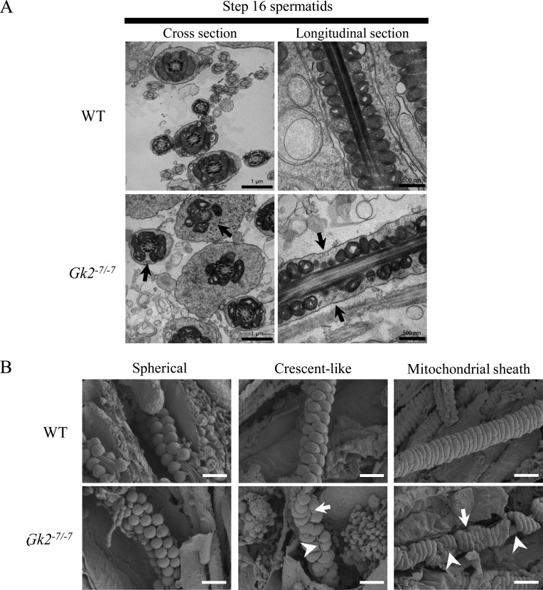
Gk2 deficient spermatids show aberrant mitochondrial sheath formation caused by misalignment of crescent-like mitochondria. (A) Ultrastructural images of step 16 spermatids (stage VII) analyzed by transmission electron microscopy (TEM). Arrows indicate the midpiece not surrounded by mitochondria. Scale bars of cross section and longitudinal section are 1 µm and 500 nm, respectively. (B) Mitochondrial sheath development during spermatogenesis observed by scanning electron microscopy (SEM). During spermatogenesis, spherical mitochondria align around flagellum (left), and change their shape to crescent-like mitochondria to surround the flagellum (middle), and then the mitochondria continue to elongate to form mitochondrial sheath (right). Arrows show breaks of aligned mitochondria. Arrowheads show exposed outer dense fiber. Scale bars are 1 µm.

