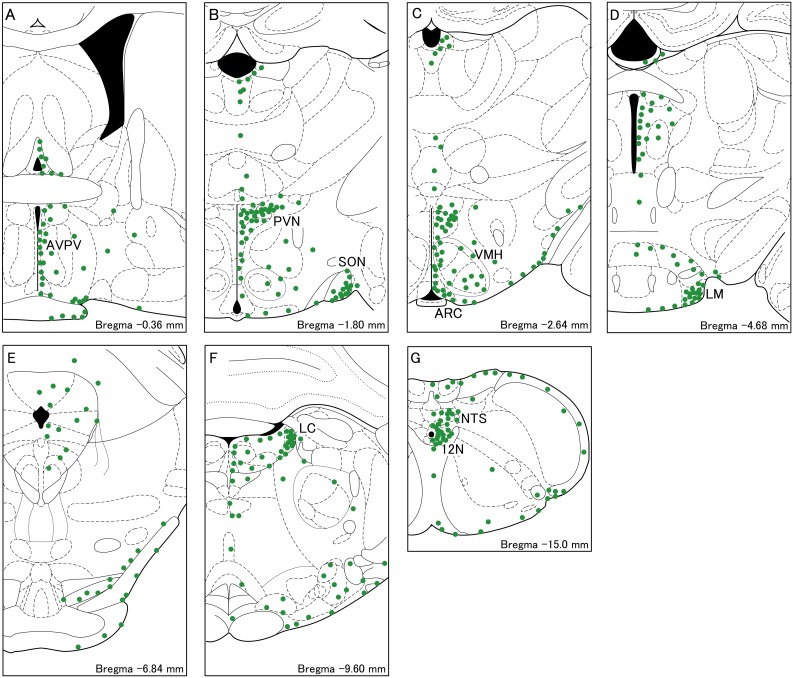
Fig. 2.
Schematic illustrations of WGA-immunopositive cells in the brainstem frontal sections of a representative female rat injected with WGA into the 4V. The distribution of the cell bodies containing WGA (green dots) are shown in A–G. A dot indicates 20 cells. AVPV, the anteroventral-periventricular nucleus; PVN, the paraventricular nucleus; SON, supraoptic nucleus; ARC, the arcuate nucleus; VMH, the ventromedial hypothalamus; LM, the lateral-mammillary nucleus; LC, the locus coeruleus;12N, hypoglossal nucleus; NTS, the solitary tract nucleus.

