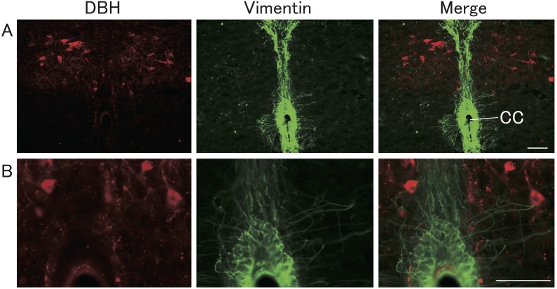
Fig. 7.
Localization of vimentin and DBH immunoreactivities in the brainstem. Representative photomicrographs of sections showing DBH-immunopositive (red) and vimentin-immunopositive (green) cells and fibers in the brainstem including the A2 region of NTS and the central canal (A). High magnification of photomicrographs is shown in B. Scale bars = 50 μm.

