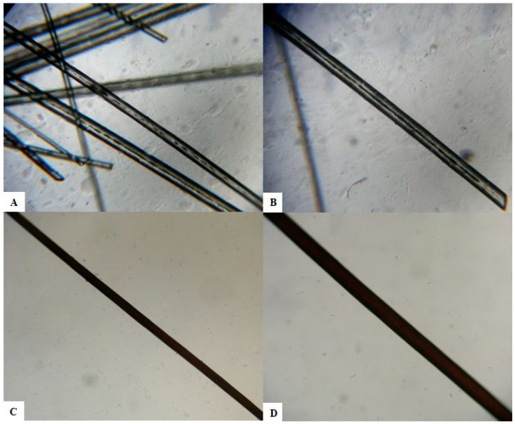
Figure 2.
Light microscopic examination of hair shafts of the proband (A,B) and of a healthy control (C,D). Giant uneven melanin granules in the medullar zone of the hair’s proband at light microscopy (A) 40× magnification; (B) 100× magnification. Aspect on light microscopy of a black hair of a healthy Caucasian control. The pigment is homogeneous and evenly distributed in the hair shaft. (C) 40× magnification; (D) 100× magnification.

