
Fig. 4.
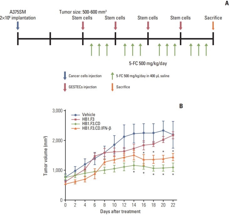
Changes in tumor volume of melanoma A375SM cell line following human neural stem cell (hNSC) treatments. A xenograft model was created by implanting A375SM cells (2×10⁶ cells) into female athymic mice. (A) CM-DiI pre-stained hNSCs (4×10⁶ cells) were injected to adjacent tumor masses after the tumor mass reached between 500 and 600 mm3 . Intraperitoneal injection of 5-fluorocytosine (5-FC; 500 mg/kg/day) was implemented for 3 days after 2 days of hNSC injection. (B) The measurement of tumor volumes was carried out for 3 weeks and calculated as length×width×height×0.5236 (mm3 ). The changes in tumor after treatment with hNSCs in the presence of 5-FC were displayed in a graph. Data were represented as mean±standard error of mean. *p < 0.05 vs. 5-FC treatment alone.