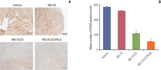
Fig. 6.
Immunohistochemistry analysis of proliferating cell nuclear antigen (PCNA) in a tumor mass in melanoma xenograft mouse model. The expression of PCNA in tumor masses after immunohistochemical analysis was shown. Tumor masses were taken from every mouse. A 10% formalin fixation, paraffin embedding, and 5-μm sectioning using a microtome were performed. (A) A primary antibody specific for PCNA was used and the data were analyzed using a microscope. (B) The mean number of PCNA positive nuclei of each section was quantified and shown in the graph. Data are represented as mean±standard error of mean. *p < 0.05 vs. vehicle (5-fluorocytosine).

