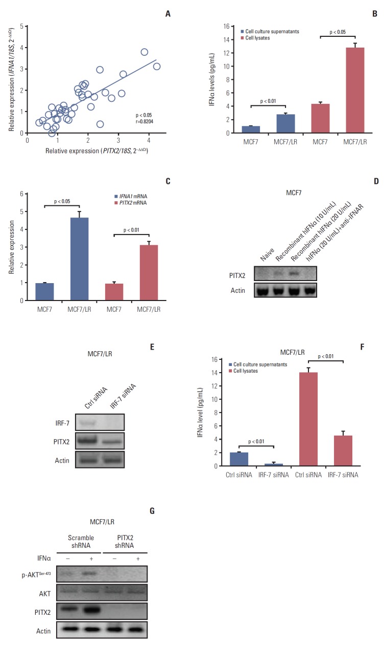
Fig. 4.
Activation of interferon α (IFNα) signaling pathway stimulates paired-like homeodomain transcription factor 2 (PITX2) expression in letrozole-resistant breast cancer (BCa) cells. (A) PITX2 and IFNA1 expression levels in primary BCa tissues (n=24) and recurrent BCa tissues (n=20) were determined using quantitative real-time polymerase chain reaction (RT-qPCR). Fold change was determined for each sample relative to the internal control gene 18S. Each value is a mean±standard error of mean from three experiments. The correlation between PITX2 and IFNA1 expression levels were the analyzed using Pearson chi-square test. (B) Enzyme-linked immunosorbent assay (ELISA) analysis of baseline expression of IFNα in cell lysates and culture supernatants in different BCa cells. (C) Relative expression levels of PITX2 and IFNA1 mRNA were assayed using RT-qPCR in different BCa cells. (D) MCF7 cells were challenged for 24 hours with a gradual concentration of recombinant hIFNα protein, in the presence or absence of the pretreatment with 5 μg/mL of anti-IFNAR neutralizing antibody for 4 hours. PITX2 expression was then determined using Western blotting. Actin served as the loading control. (E) MCF7/LR cells were transiently transfected with IRF-7 siRNA or Ctrl siRNA using Lipofectamine 3000. Forty-eight hours later, cells were harvested and subjected to Western blotting analysis of IRF-7 and PITX2 expression. (F) ELISA analysis of baseline expression of IFNα in cell lysates and culture supernatants in MCF7/LR cells with different transfections. (G) MCF7/LR cells with different transfections were challenged with 20 U/mL of recombinant hIFNα protein for 24 hours, followed by Western blotting analysis of p-AKTSer-473, AKT and PITX2 expression.

