
Fig. 4.
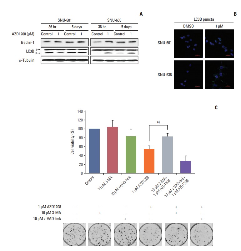
Induction of cell death by AZD1208 through stimulation of autophagy. (A) Cells were treated with dimethyl sulfoxide (DMSO; control) or 1 μM AZD1208 for 36 or 120 hours. The expression levels of light chain 3B (LC3B) and Beclin-1 were measured by western blot analysis. α-Tubulin was used as a loading control. (B) SNU-601 and SNU-638 cells transfected with GFP-LC3B were treated with 1 μM AZD1208 for 5 days. Confocal microscopy was used to observe the signals corresponding to LC3B expression (green fluorescence). DNA was counterstained with DAPI (blue). The merged images represent overlapping signals of the two channels. (C) SNU-638 cells were pre-treated with the autophagy inhibitor 3-methyladenine (3-MA; 10 μM) or caspase-3 inhibitor z-VAD-fmk (10 μM) for 24 hours. Next, the cells were treated with 1 μM AZD1208 every 3 days for 14 days. The percentages of surviving cells were calculated by counting the number of colonies and are presented in a bar graph with standard error bars (n=3). a)p=0.008.