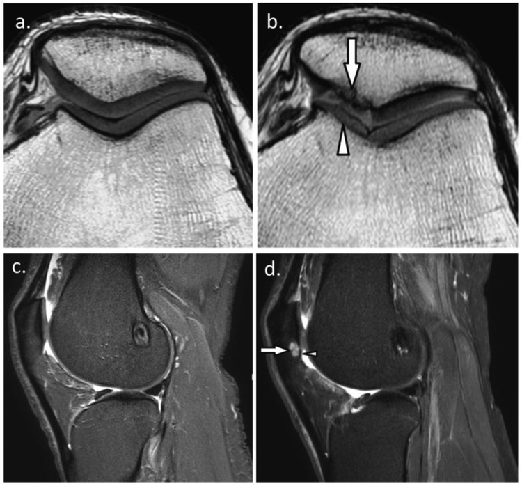
Figure 2.
a. 26-year-old man with ACL reconstruction. Baseline sagittal short-tau inversion recovery (STIR) MRI shows normal cartilage of the patellofemoral joint and absence of bone marrow lesion, and b. Follow-up MRI at the same level shows new small superficial focal cartilage defect at the inferior pole of the patella (arrowhead) with associated subchondral bone marrow lesion (arrow); c. 31-year-old man with ACL reconstruction. Baseline axial proton density-weighted MRI shows normal patellofemoral cartilage, and d. Follow-up MRI at the same level shows diffuse superficial thinning of the medial trochlear cartilage (arrowhead) and large full thickness cartilage loss at the medial patella (arrow) with denuded bone area.

