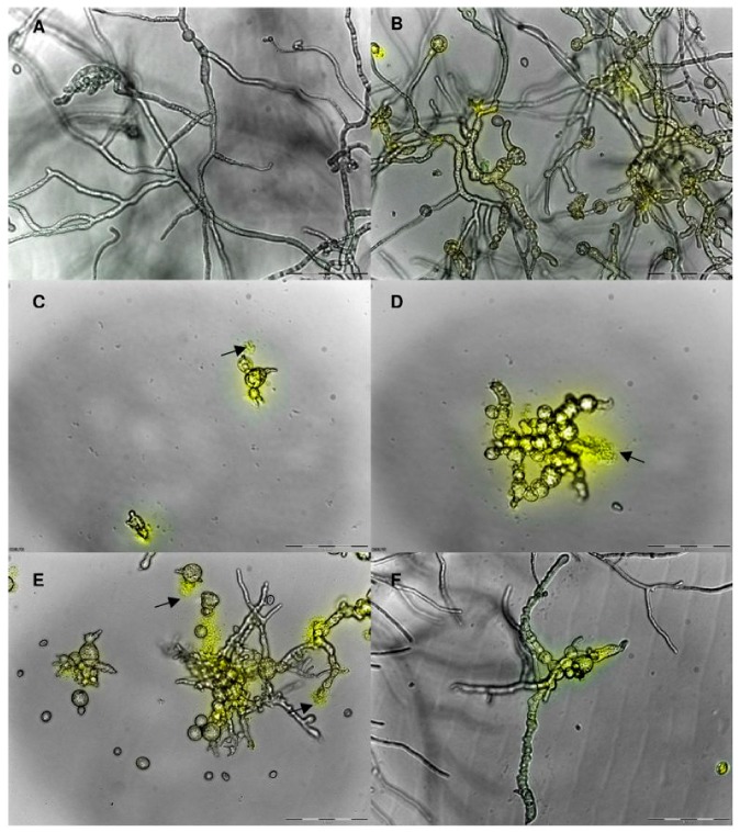
Figure 4.
Combined overlay of the light microscopical analysis at 20× magnification and the cell permeabilization assay conducted on B. cinerea grown in the presence of Hc-AFPs for 48 h at 23 °C. (A) Control, (B) Hc-AFP1 25 μg/mL, (C and D) Hc-AFP2 15 μg/mL, (E) Hc-AFP3 25 μg/mL, (F) Hc-AFP4 18 μg/mL. The yellow fluorescence indicates a compromised membrane and the black arrows indicate structures that are leaking their cellular content into the surrounding medium. Adapted from De Beer and Vivier [31], an open-access article distributed under the terms of the Creative Commons Attribution License (http://creativecommons.org/licenses/by/2.0). Copyright (2011) the authors, licensee BioMed Central Ltd.

