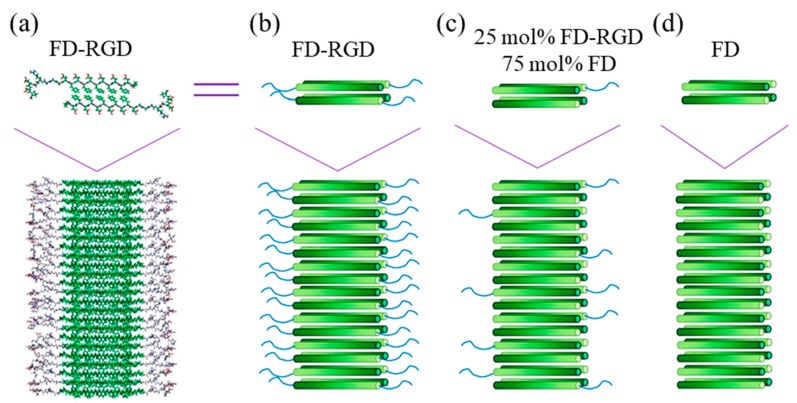
Figure 6.
(a) Scheme showing a molecular model of Pro-DFDFDFDFDFDGGGRGDS-Pro (FD-RGD) assemblies) and (b–d) cylinder representations with a gradient color from the N- to C-termini (dark to bright, respectively). The β-strand conformation positions the hydrophobic Phe side chains from both layers facing each other, while the hydrophilic side chains point to the surrounding aqueous phase. The top schemes show four peptides arranged in a bilayer that constitutes the fibril shown in the bottom scheme. Reproduced from [120].

