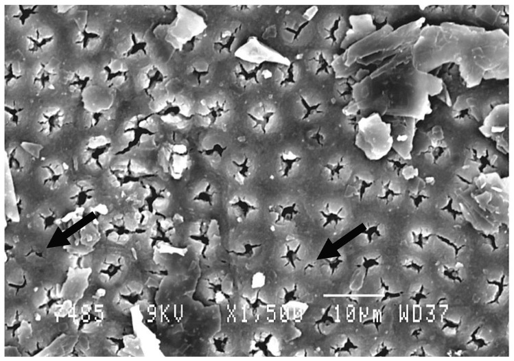
Figure 4.
Scanning electron microscopic (SEM) views of treated dentin by diode laser (980 nm) at 1 W. A narrowing of dentinal tubules can be noted. Only a few tubules are completely obliterated. Graphite particles still exist on the dentinal surface (not disintegrated by the laser beam). Magnification: 1500×.

