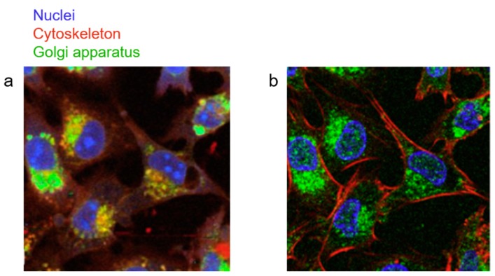
Figure 2.
Comparison of Raman-based, artificial IF and fluorescence imaging. (a) A Raman-based, artificial IF image of cultured human glioma cells with false color assignments showing the nuclei in blue, cytoskeleton in red, and Golgi apparatus in green. (b) An immunofluorescent image of the same cells staining the nuclei (DAPI, blue), cytoskeleton (rhodamine-conjugated phalloidin, red), and Golgi apparatus (anti-Syntaxin-6-antibody, green). Reproduced with permission from [63], Copyright Elsevier, 2012.

