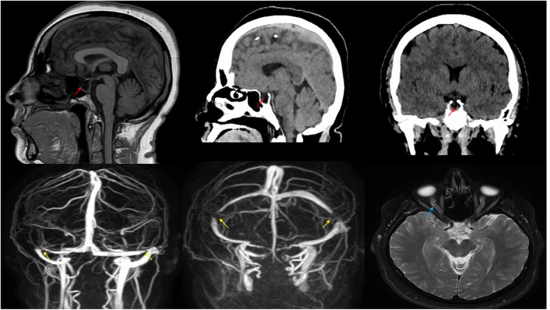
Fig. 1.

Top: A computed tomography scan of the head in the top middle picture showed pontomedullary hypodensity, and a partially empty sella turcica, noting the top first one on the left is brain MRI (red arrows). Bottom: Brain magnetic resonance imaging with magnetic resonance venography revealed a stenosis in the lateral aspect of the transverse sinus (yellow arrows), and perioptic nerve sheath distention which is consistent with papilledema (blue arrow)
