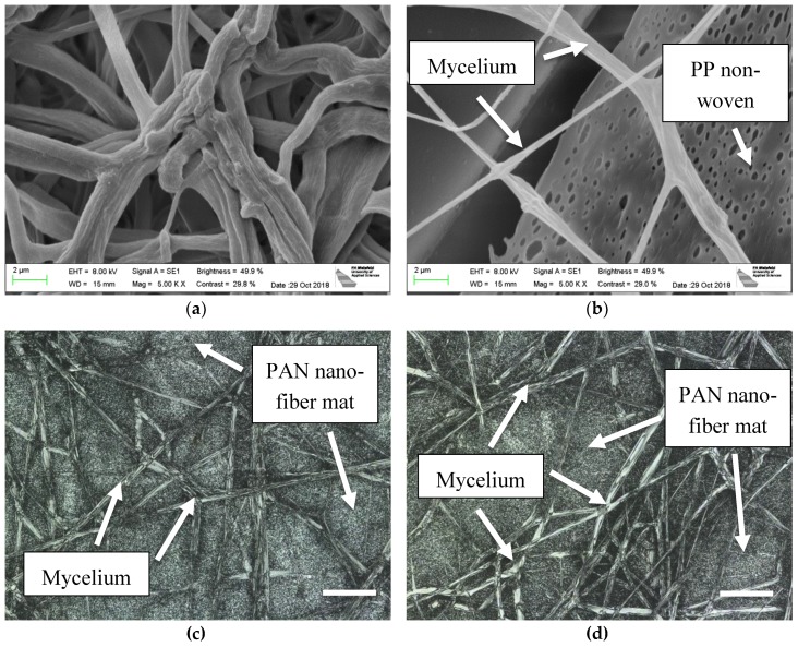
Figure 3.
SEM images of oyster mushroom mycelium (a) grown on agar; (b) grown between a PAN nanofiber mat and the nonwoven PP used as a typical support for electrospinning; scale bars indicate 2 µm; CLSM images of (c) mycelium on PAN nanofiber mat; (d) mycelium under PAN nanofiber mat, being grown through it. Scale bars indicate 20 µm.

