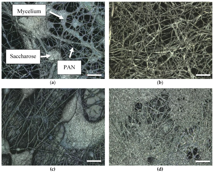
Figure 6.
CLSM images: (a) oyster mushroom mycelium (thick fibers) on PAN/saccharose nanofiber mat (thin PAN fibers and saccharose agglomerations); (b) oyster mushroom mycelium on PAN/poloxamer nanofiber mat (the latter not visible here); (c) and (d) PAN/saccharose nanofiber mats after watering. Scale bars indicate 20 µm.

