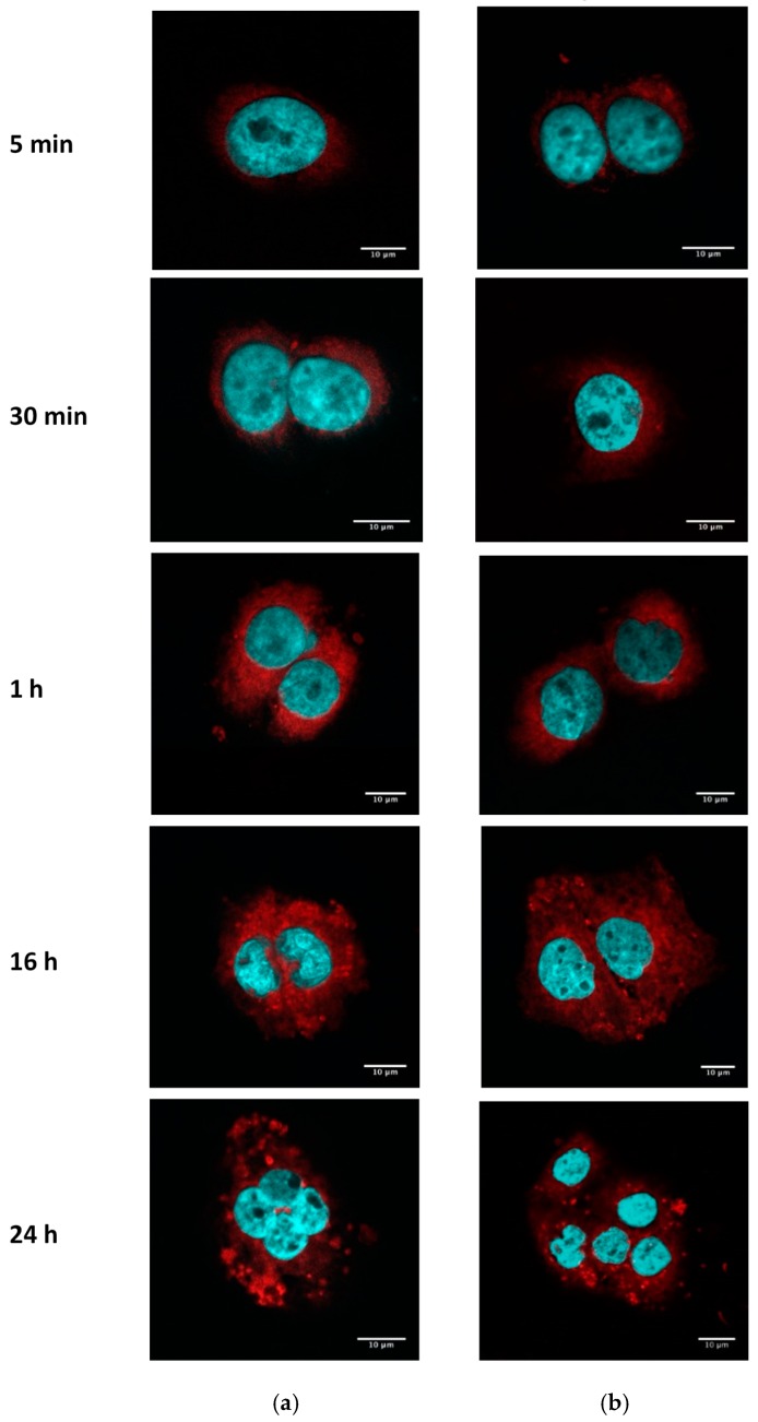
Figure 5.
Internalization of fSLN and fPEG–SLN in SCC-25. SCC-25 cells were seeded in cover glasses at 2 × 10⁴ cells/well. The next day, cells were washed and incubated with 0.2 mg/mL of fPEG–SLN (a) or fSLN (b) suspensions. Fluorescence intensity of R18 (red) was analyzed by confocal microscopy and nuclei were stained with DAPI (blue). Scale bar: 10 µm.

