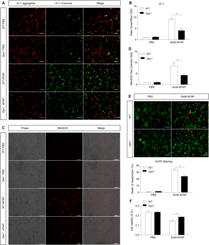
Fig. 5.
In vitro protection against APAP-induced mitochondrial dysfunction in primary hepatocytes with Opn depletion. Isolated primary WT and Opn−/− hepatocytes were starved and subjected to APAP treatment. a Representative images of mitochondrial membrane potentials in primary hepatocytes 6 h after APAP treatment determined using JC-1 (origin magnification ×100); b integrated optical density (IOD) quantification of the ratio of green to red fluorescent intensity (n = 3). c Representative images of mitochondrial ROS in primary hepatocytes 6 h after APAP treatment determined with the MitoSOX Red probe (origin magnification ×100). d Quantification of MitoSOX Red intensity (n = 3). e Representative images of hepatocyte death 12 h after APAP treatment determined with acridine orange and propidium iodide (AO/PI) staining (origin magnification ×100). Quantification of the percentages of PI-stained dead hepatocytes (n = 3). f Hepatocyte viability 12 h after APAP treatment determined using the MTT assay (n = 5). Data are shown as the means ± SEM, *P < 0.05

