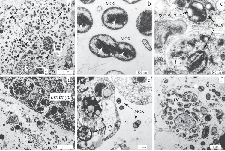
Fig. 3.
Transmission electron microscopy images of H. (S.) methanophila. a Symbiotic methane-oxidizing bacteria (MOX) are abundant in the mesohyl, particularly in regions close to the choanocyte chambers (chambers not visible in image), sp = sponge cells, n = nucleus. b High-resolution image of white box labeled b from panel (a), showing intracellular membranes (arrows) typical for MOX. The MOX at the lower half of the image are dividing; bc = bacteria with a different morphology than the MOX symbionts. c MOX symbiont in a lysosome of an amebocyte, l = lysosome. d Mesohyl with an embryo surrounded by symbiotic MOX, and a bacteriocyte containing bacteria with a different morphology than the symbiotic MOX (bc). e MOX symbionts within an embryo. he = heterogeneous yolk. f A bacteriocyte containing bacteria with a different morphology than the MOX symbionts. bc = bacteria; n = nucleus

