
Figure 3.
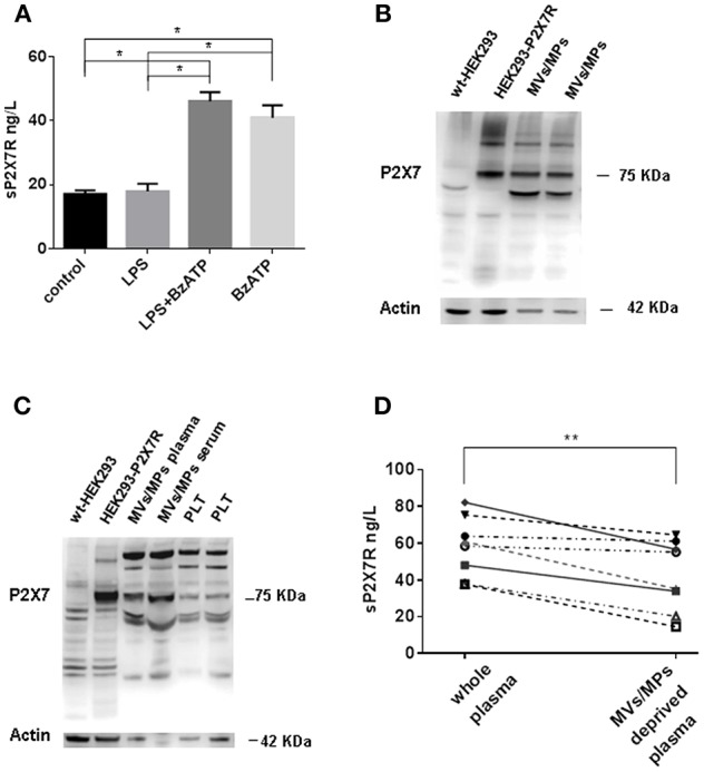
sP2X7R is shed in association with MVs/MPs by P2X7R stimulation. (A) Monocyte-derived macrophages were isolated from blood samples of 4 healthy control subjects as described in Methods and incubated in 10% FBS-supplemented RPMI under the following conditions: (1) no additions (control) for 5 h; (2) 1 μg/ml LPS for 4 h; (3) 1 μg/ml LPS for 4 h, followed by 300 μM BzATP for 1 h; (4) no additions for 4 h, followed by 300 μM BzATP for 1 h. Data are means ± SE of data from 4 separate experiments. Only significant differences are shown in the graph. Kruskal-Wallis test p = 0.0008. Mann Whitney test between groups: *p < 0.05. (B) MVs/MPs were isolated from two plasma samples by centrifugation at 2200xg followed by 14000xg. Ten μg of protein were loaded on the SDS-PAGE gel. (C) MVs/MPs were isolated from one plasma sample and from one serum sample by centrifugation at 2200xg followed by 100000xg ultracentrifugation. Platelets (PLT) were isolated from plasma by centrifugation at 2200xg. See Material and Methods for additional details. Ten μg of proteins were loaded on the SDS-PAGE gel. (D) ELISA analysis of sP2X7R content of plasma samples from 8 subjects before and after depletion of MVs/MPs by ultracentrifugation at 14000× g. Wilcoxon matched paired t-test: p = 0.0078.