
FIGURE 2.
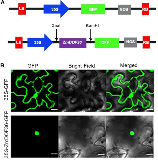
Subcellular localization of ZmDOF36. (A) Schematic diagram of the DNA construct used for ZmDOF36 subcellular localization. LB, T-DNA left border; 35S, cauliflower mosaic virus 35S promoter; GFP, green fluorescent protein; NOS, nopaline synthase gene terminator; RB, T-DNA right border. (B) The 35S::ZmDOF36-GFP fusion protein and 35S::GFP DNA constructs were transiently expressed separately in Nicotiana benthamiana leaf epidermal cells and visualized with a confocal laser scanning microscope. Scale bar = 20 μm.