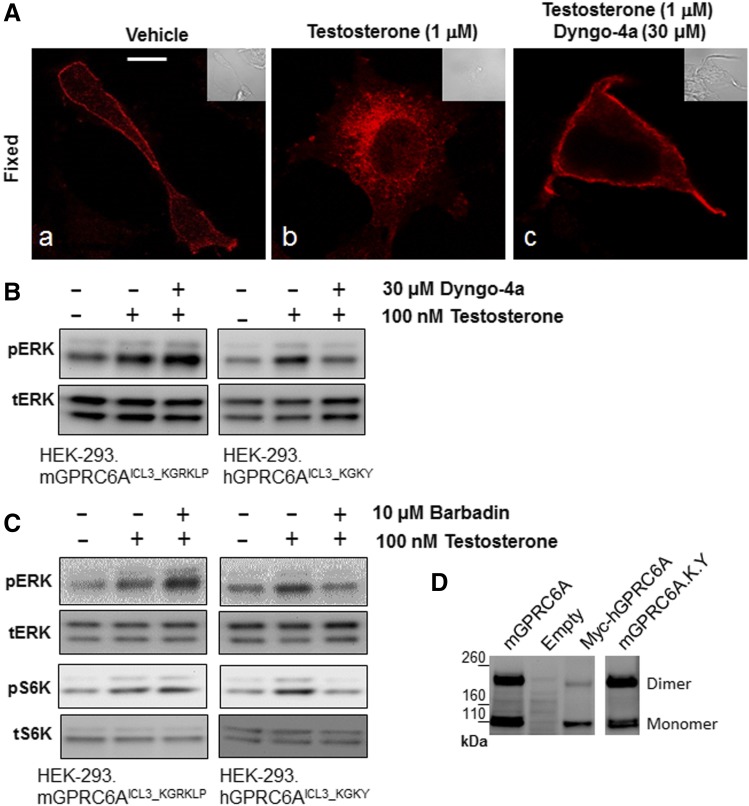
Fig. 5.
Comparison of intracellular function of mouse and human GPRC6A in HEK-293 cells. (A) Dyngo-4a inhibited GPRC6A ligand; testosterone induced receptor internalization. The slides were pretreated with diluent or 30 µM Dyngo-4a for 30 minutes and then exposed to 1 µM testosterone for 30 minutes and fixed (images b and c, respectively). Scale bar, 5 μm. (B and C) Comparison of Dyngo-4a and Barbadin regulated GPRC6A-mediated ligand; testosterone induced ERK and S6K phosphorylation in HEK-293 cells stably transfected with WT mouse and human GPRC6A (mGPRC6AICL3_KGRKLP and hGPRC6AICL3_KGKY) cDNAs. Cells were preincubated with vehicle, Dyngo-4a (30 μM (Ab120689; Abcam), or Barbadin (10 μM) (Axon2774; Axon Medchem) for 20 minutes after testosterone treatment. (D) Comparison of dimerization of the various GPRC6As in HEK-293 cells, WT mouse GPRC6A (mGPRC6AICL3_KGRKLP) vs. humanized mouse GPRC6A (mGPRC6AICL3_KGRKLP) or human GPRC6A (hGPRC6AICL3_KGKY), analyzed by western blots: 16 μg of each plasmid cDNA was transfected into HEK-293 cells (1.5 × 10⁶ cells/100 mm dish) and 100 μg of each total protein sample was loaded on 3%–8% SDS-PAGE gels. Anti-Myc antibody was used in the western blot analysis. These images are representative of images derived from three separate experiments.

