FIGURE 2.

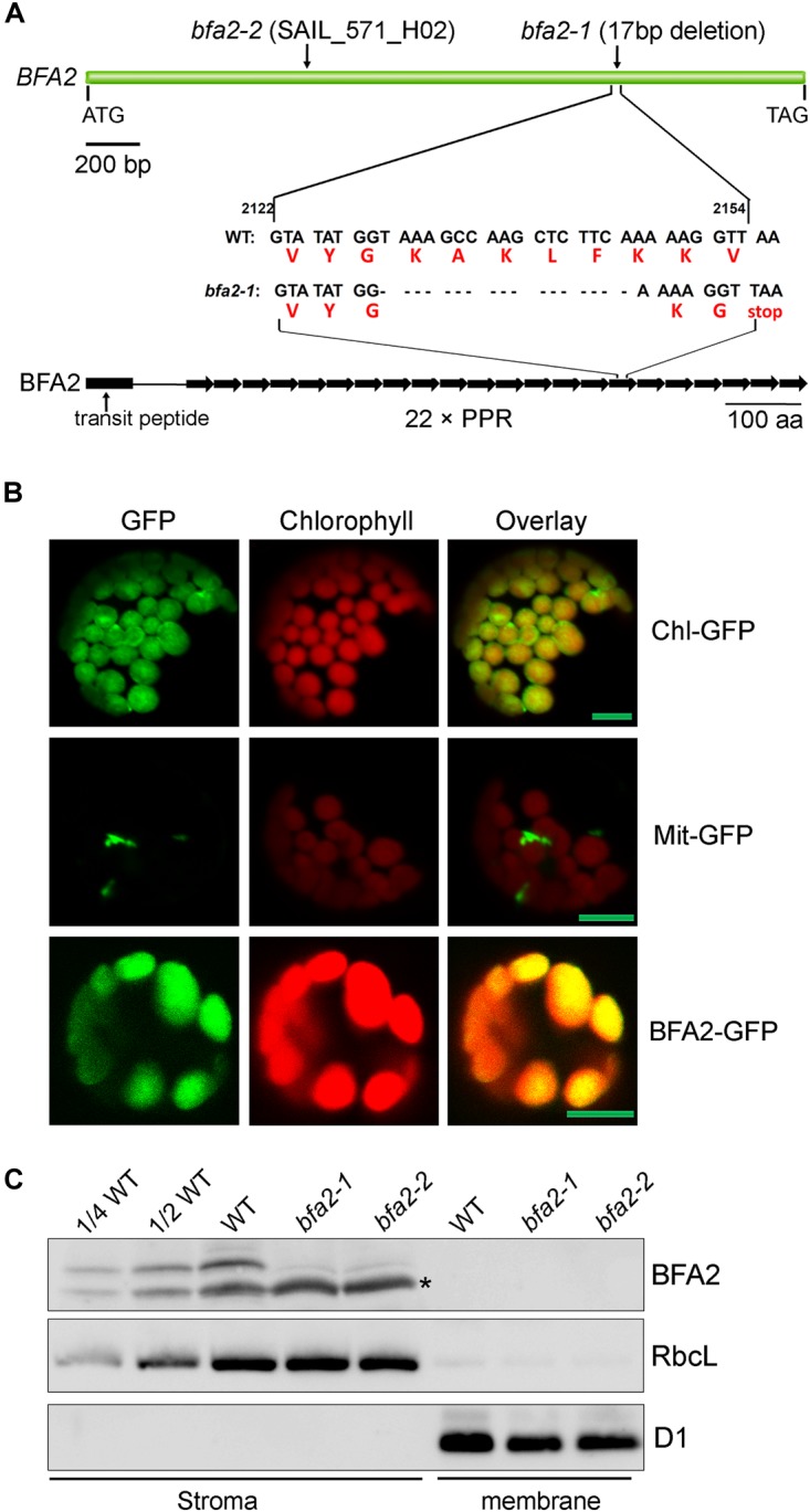
Characterization of the BFA2 protein. (A) Schematic representation of BFA2 gene (Top panel) and BFA2 protein (bottom panel). Positions for nucleotide deletion in baf2-1 and T-DNA insertion in bfa2-2 are indicated. Each right arrow represents one PPR domain. The 17-nucleotide deletion results in a premature stop codon at the position of the 16th PPR motif in BFA2. (B) Subcellular localization of BFA2 by GFP assay. The first 200 amino acids of BFA2 were fused with GFP (BFA2-GFP) and expressed in Arabidopsis protoplasts. The signal of GFP was visualized using a confocal laser scanning microscope. Chl-GFP and Mit-GFP represent chloroplast and mitochondrial controls, respectively. Bars = 5 μm. (C) Immunolocalization of BFA2. Intact chloroplasts isolated from WT and bfa2 mutants were fractionated into stromal and membrane fractions. Proteins were separated by SDS-PAGE and immunodetected with antibodies against BFA2, RbcL, and D1. The series of WT dilutions is indicated. A major nonspecific band detected in the stromal fractions with BFA2 antibody is indicated by an asterisk. A weak band above the major nonspecific band detected in bfa2 stroma also appears to be nonspecific.
