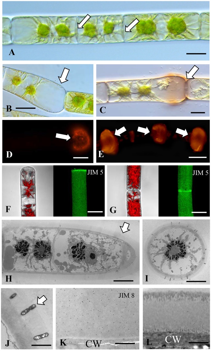
FIGURE 4.
Zygnema: (A) Cross walls of the unbranched filament (arrows). Bar7 μm. (B) Cell wall (arrow) at swelling of cell at a fragmentation site (arrow). Bar 12 μm. (C) ß-glucosyl-Yariv labels the wall and sheath of the swollen zone (arrow). Bar 12 μm. (D) JIM13 labeling of ECM at the swollen fragmentation zone (arrow). Bar 15 μm. (E) JIM13 labeling of “clouds” of ECM at the cross walls of filaments (arrows). Bar 15 μm. (F) JIM5 labeling (right) of terminal cell and corresponding bright field image with chloroplast autofluoescence (left). (G) JIM5 labeling of two connected cells, notice the two rings in the connection zone (arrow). Bars (F,G) 20 μm. (H) TEM image of young filament with broad electron translucent ECM layer surrounding the cells (arrow). (I) Cross section through filament. Bars (H,I) 20 μm. (J) Bacteria in the ECM layer of a young filament (arrow). (K) JIM8 immuno-gold labeling of the ECM layer outside the cell wall (CW). (L) ECM of older cell, notice the fibrillary structures outside the CW and at the periphery. Bars (J–L) 2 μm.

