
FIGURE 5.
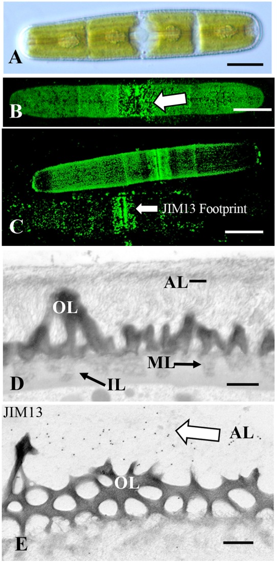
Penium: (A) DIC image of Penium. Bar 12 μm. (B) JIM13 labeling of the outer surface of the cell wall (arrow). Bar 15 μm. (C) JIM13 footprint (arrow) of adhesive material left on substrate after removal of cells. Bars 15 μm. (D) TEM image of the cell wall revealing the pectin-rich outer wall (OL) that connects to a medial layer (ML) that is embedded in an inner layer (IL). External to the outer layer is an adhesive layer (AL) of fine fibrils (arrows). Bar 300 nm. (E) JIM13 labeling (arrow) of the outer adhesive layer (AL) of the wall. Bar 250 nm.