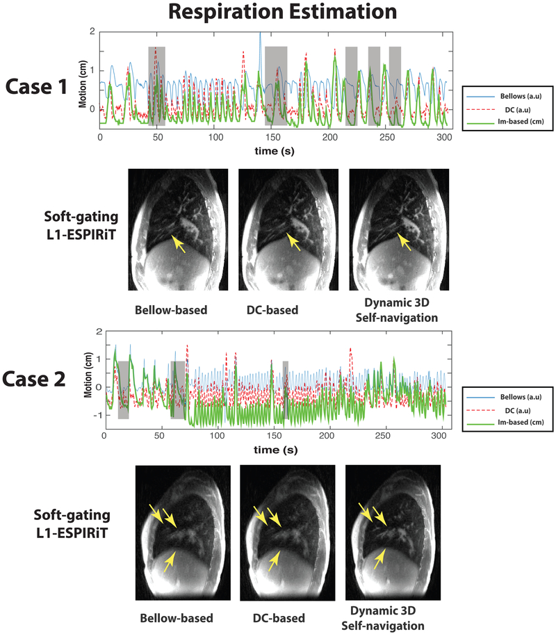
FIG. 3.
Comparison of different respiration estimation methods in CF patient case 1 with a mildly irregular breathing pattern and CF patient case 2 with a strongly irregular breathing pattern: In plots, (blue) respiration motion estimated by the respiratory bellows belt (“bellows”); (red dashed) respiration motion derived from the center of k-space (“DC”); (green) respiration SI motion derived from the dynamic 3D navigator (“Im-based”). Note that the only dynamic 3D self-navigation signal is quantitative (units of cm) whereas the DC-based and bellows are only relative measurements. Gray shaded boxes highlighted the time points where bellow signal/DC-based self-navigation differ a lot with dynamic 3D navigator. Comparisons of soft-gating L1-ESPIRiT reconstructions based on corresponding respiration estimations are shown below the plots. These reconstructions are based on a soft weighting window centered at the end of expiration. The arrows indicate vessels with varying conspicuity based on different respiration estimations.

